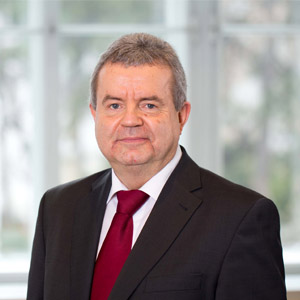
Keynote Speaker

Prof. Siegfried Trattnig
High Field MR Center, Department of Biomedical Imaging and Image Guided Therapy, Department of Neurology, Medical University of Vienna, AustriaSpeech Title: Ultrahigh field MR (7 Tesla) in the advanced diagnosis of Multiple Sclerosis
Abstract:
- Ultrahigh field (7 Tesla) due to higher spatial resolution and higher sensitivity to susceptibility effects (SWI) improves Central Vein Sign (CVS) in MS lesions at 3T: 45%, 7T: 87% visibility. The CVS is an excellent differential diagnostic criterium between MS lesion and lesions of vascular origin and MS mimics.
- A subgroup of MS lesions (40%) show iron rim lesions (IRL). IRLs are composed of proinflammatory iron containing microglia cells and macrophages and are present in all MS stages with a peak in late RRMS and early SPMS phase. IRL gradually increase in size and fuse with neighbouring IRL within years. Whereas non-IRL shrink over time. Lesions with iron rim are slowly expanding lesions. Therefore IRL are suggested to label a subset of chronic active lesions and are worse in prognosis for IRL comared to non-IRL. IRL correlate with clinical disability. Only one-third of IRL seen at 7T can be seen at 3T. High-resolution MRSI at 7T in Multiple Sclerosis
- 7 Tesla allows higher spectral resolution and high-resolution spectroscopic imaging (MRSI) in shorter scan times compared to 3T. In normal-appearing white matter neurochemical changes can already be seen with Myo- Inositol (mIns) increase to be an earlier imaging biomarker for neuroinflammation compared to conventional MR. Mean mIns/NAA ratios correlated significantly with Clinical Disability measured with the EDSS score. In summary MRSI at 7T allows detection of early lesion development and their activity, evaluation of disease progression and sensitive monitoring under therapy as well as monitoring of new treatment trials.
Biography: Univ. Prof. Dr. Siegfried Trattnig graduated from the University of Vienna Medical School in 1985. He trained in Radiology and subsequently served as Assistant Medical Director and Acting Medical Director for the Section of Neuroradiology in the Department of Radiology, Medical University of Vienna. He was appointed as an Associate Professor in Radiology 1993 becoming the Acting Medical Director at the Clinical Magnetic Resonance Institute at the University of Vienna. Since 2003 Prof Trattnig has the position of the Medical Director of the Centre of Excellence in high-field MR at the Medical University of Vienna. In 2010 he was appointed as a full Professor for Radiology with special focus on High field MR. Prof. Trattnig has pioneered the field of multi parametric or biochemical MR imaging of cartilage and in particular cartilage repair. Together with Stefan Marlovits he has developed the semiquantitative MR Observation of Cartilage Repair Tissue Score (MOCART) 1.0 and 2.0 which is now widely accepted by the orthopedic community and the authorities such as the FDA. He is currently the lead researcher on the clinical 7T & 3T projects at the Medical University in Vienna. Based on the results of clinical comparison studies between 3 and 7T his Center of Excellence for High Field MR was appointed as the international Reference Center for 7 Tesla by Siemens Healthcare, the leader in the ultra-high field MR.
He is or has been editorial board member of 8 scientific journals, member of 35 committees and working groups within the ISMRM, ESR, ESMRMB and the ICRS among the Executive Board member of the ESMRMB, member of the ESR Research Committee Board and Chairperson of the ESR Subcommittee Imaging Biomarker. He is an author of 648 articles in peer reviewed scientific journals and contributed to 25 scientific books. His h-Index is 79. Additionally he has held 26 peer reviewed scientific grants with a total of funding money of 14.5 Mio €, received 12 scientific awards and is a reviewer for 34 scientific journals. In 2019 he was awarded with the Senior Fellowship of the ESMRMB and in 2021 with the Senior Fellowship of the ISMRM.
